Myelin, the fatty insulation wrapping nerve fibers throughout your brain and spinal cord, depends on a specific class of lipids that most people have never heard of: plasmalogens. These ether-bonded phospholipids make up a substantial fraction of myelin’s total lipid mass, and when plasmalogen levels drop, myelin integrity suffers. Understanding the relationship between plasmalogens and myelin offers a window into why nerve sheath health matters so much for cognition, movement, and overall neurological function.
Research over the past five decades has mapped the lipid architecture of myelin in increasing detail, revealing that plasmalogens are not passive structural fillers but active contributors to membrane stability and signaling. More recent work has identified the enzymes and trace mineral cofactors required to produce these lipids, connecting nutritional status to myelination in ways that were previously only theoretical. This article walks through the evidence linking plasmalogens to myelin nerve sheath integrity and considers where trace minerals like selenium and iron fit into the picture.
Key Takeaways
- Plasmalogens are ether-bonded phospholipids that constitute a major fraction of myelin and are essential for nerve sheath structure and stability [1].
- Genetic disruption of plasmalogen synthesis enzymes—including Far1 and selenoprotein I—causes hypomyelination and neurological deficits in animal models [2] [3].
- Selenium and iron serve as cofactors for oligodendrocyte function and ether lipid production, linking trace mineral status to myelination capacity [4].
- Shilajit contains trace selenium and iron, but no clinical evidence directly connects shilajit supplementation to plasmalogen levels or myelin health.
- Myelin lipid composition is sensitive to genetic, nutritional, toxic, and metabolic disruptions across the lifespan, making the ether lipid pathway an area of growing research interest.
What Are Plasmalogens and Why Do They Matter for Myelin?
Plasmalogens are a subclass of glycerophospholipids defined by a vinyl-ether bond at the sn-1 position of the glycerol backbone, rather than the typical ester bond found in most membrane phospholipids. This structural difference gives plasmalogens unique biophysical properties: they pack more tightly in membranes, influence membrane fluidity, and can act as endogenous antioxidants by scavenging reactive oxygen species at the vinyl-ether linkage.
In the central nervous system, plasmalogens are especially concentrated in myelin sheaths. Early biochemical studies of human brain tissue established that ethanolamine plasmalogens account for a large share of total myelin phospholipids [1]. An early analysis of myelin from multiple sclerosis patients found no detectable deficit in plasmalogens [5]. Genetic models suggest a functional requirement: myelin needs plasmalogens to maintain its characteristic tightly compacted, multilayered structure.
Axonal membranes also contain plasmalogens, though in different proportions than myelin itself. Analysis of axolemma-enriched fractions from human brains revealed distinct lipid profiles between axonal and myelin membranes, with plasmalogens present in both compartments but particularly enriched in myelin [6]. This distribution pattern suggests that plasmalogens serve specialized roles in the insulating sheath that surrounds nerve fibers.
How Plasmalogen Deficiency Disrupts Myelination
The clearest evidence for the functional importance of plasmalogens in myelination comes from genetic models where ether lipid synthesis is impaired. A recent study on mice lacking Far1, an acyl-CoA reductase essential for the first step of ether lipid biosynthesis, demonstrated that loss of this enzyme leads to severely reduced plasmalogen levels and pronounced hypomyelination in the brain [2]. These animals showed thinner myelin sheaths, fewer myelinated axons, and measurable neurological impairments, establishing a direct causal link between ether lipid production and myelin formation.
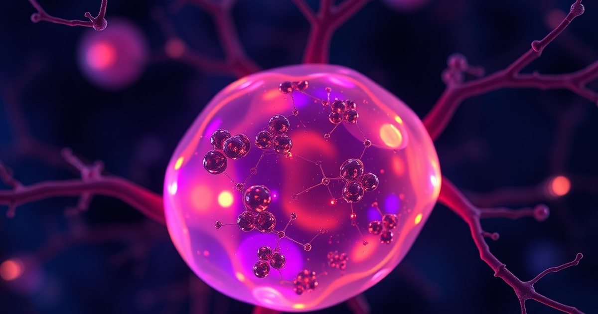
Similarly, research on selenoprotein I, an enzyme involved in ether lipid homeostasis, found that its deletion in mice caused dramatic reductions in plasmalogens and corresponding defects in myelination [3]. The affected animals displayed tremors, motor deficits, and reduced nerve conduction, all consistent with inadequate myelin insulation. Naturally occurring neurological mutant mice have also been surveyed for myelin lipid abnormalities, with systematic analysis finding alterations in several other lipid classes while plasmalogen levels were normal in all of the mutants [7].

Inherited metabolic disorders in humans tell a similar story: in Refsum’s disease, where abnormal lipid metabolism disrupts the nervous system, detailed analysis of brain lipids revealed significant changes in plasmalogen content alongside broader lipid abnormalities [8]. Across genetic and disease models, the pattern is consistent—when the ether lipid pathway is compromised, myelin quality suffers.
The Selenium and Iron Connection to Ether Lipid Synthesis
Producing plasmalogens is not just a matter of having the right enzymes. Certain trace minerals serve as essential cofactors in the biosynthetic and protective pathways that keep ether lipid levels adequate for myelination. Two minerals with well-documented roles are selenium and iron.
Selenium is incorporated into selenoproteins, a family of enzymes with diverse functions in antioxidant defense and lipid metabolism. Selenoprotein I directly participates in ether lipid synthesis, and its activity depends on adequate selenium availability [3]. A comprehensive recent review examined how both iron and selenium intersect at critical points in oligodendrocyte biology—the cells responsible for producing myelin in the central nervous system [4]. Iron is required for oligodendrocyte differentiation and for the energy-intensive process of synthesizing large quantities of myelin membrane. Selenium, through selenoproteins, protects oligodendrocytes from oxidative damage during this metabolically demanding process.
When either mineral is deficient, oligodendrocyte function can be compromised, with downstream effects on plasmalogen production and myelin quality. This dual mineral requirement helps explain why nutritional deficiencies in selenium or iron during critical developmental windows can impair myelination, and it raises the question of whether supporting trace mineral status in adults could help maintain the ongoing myelin repair and turnover that continues throughout life.
Myelin Vulnerability: Toxic and Environmental Insults
Beyond genetic and nutritional factors, myelin and its plasmalogen content can be disrupted by environmental exposures. Studies on cyanide encephalopathy documented alterations in the fatty acid patterns of cerebral lipids, including changes in plasmalogen-associated fatty acids, following toxic exposure [9]. This illustrates that chemical insults to mitochondrial energy metabolism can selectively alter the lipid architecture of myelin.
More recent work has examined the effects of repeated sevoflurane exposure on developing brains. Neonatal sevoflurane exposure disrupted fatty acid metabolism and led to hypomyelination with measurable neurological consequences [10]. A parallel study tracked myelination trajectories and microglial dynamics following sevoflurane exposure, finding that the anesthetic altered the normal timeline of myelin development [11]. In a mouse model of mucopolysaccharidosis type I, analysis of the corpus callosum revealed abnormal myelin lipid composition, including shifts in the phospholipid balance that likely include plasmalogen fractions [12].
Taken together, these models reinforce that myelin is only as healthy as its lipid components, and those components are sensitive to metabolic, toxic, and environmental disruptions across the lifespan.

Where Does Shilajit Fit In? An Honest Assessment
Shilajit is a mineral-rich resinous substance traditionally used in Ayurvedic medicine. It contains trace amounts of selenium, iron, and other minerals that participate in the biological pathways described above. Because selenium is a required cofactor for selenoprotein I and other enzymes involved in ether lipid homeostasis [3], and because iron supports oligodendrocyte function and myelin synthesis [4], it is biologically plausible that adequate intake of these minerals could support the cellular machinery responsible for plasmalogen production.
However, it is important to be direct: no clinical study has measured whether shilajit supplementation affects plasmalogen levels, myelin thickness, or any marker of nerve sheath integrity in humans. The connection is indirect, running through the trace mineral cofactor pathway rather than through any demonstrated effect of shilajit on ether lipid biology. The trace mineral content of shilajit also varies considerably depending on its geographic source and processing method, making it difficult to guarantee consistent selenium or iron delivery from any given product.
Shilajit also contains fulvic acid and dibenzo-α-pyrones, compounds studied separately for effects on mitochondrial energy metabolism. Since plasmalogen biosynthesis is an energy-intensive peroxisomal process, metabolic support could in principle be relevant, but this remains speculative. The shilajit-plasmalogen connection is best treated as a hypothesis worth watching rather than a proven intervention.
🛒 Where to Buy Plasmalogen Supplements
- Prodrome Sciences ProdromeNeuro
capsules, 900 mg / 2 caps — Lab-synthesized DHA-ethanolamine plasmalogen used in Dayan Goodenowe’s research; premium-priced. - Daiwa Health Advanced Omega-3 Brain
softgels, 50 mg HSOP — Hokkaido Scallop Oil Plasmalogen softgels with natto peptides; pilot cognitive data. - REMORY Sea Squirt Plasmalogen
capsules, 30-day supply — Ascidian (sea-squirt)-derived alternative source for those avoiding scallop.
As an Amazon Associate we earn from qualifying purchases. Plasmalogen supplements vary by source (lab-synthesized vs. scallop- or sea-squirt-derived) and purity — check the form, dose, and third-party testing before buying.
A Note on the Evidence
Shilajit is not a regulated pharmaceutical product and its composition varies significantly between sources; some samples have been found to contain elevated heavy metals including lead, arsenic, and mercury, so always choose products with third-party certificates of analysis. Do not use shilajit as a substitute for medical treatment of any neurological condition, and consult your doctor before supplementing—especially if you are pregnant, nursing, taking medications, or have a diagnosed health condition.

Frequently Asked Questions
What are plasmalogens and how are they different from other lipids?
Plasmalogens are phospholipids with a vinyl-ether bond at the sn-1 position of their glycerol backbone, unlike most membrane lipids which have ester bonds. This structural difference allows plasmalogens to pack tightly in membranes, contribute to membrane fluidity regulation, and act as antioxidants. In the nervous system they are heavily concentrated in myelin sheaths [1].
Why are plasmalogens so important for myelin specifically?
Myelin is a multilayered lipid-rich membrane that insulates nerve fibers, and plasmalogens help maintain the tight packing and structural integrity of these layers. When plasmalogen synthesis is disrupted genetically, as seen in Far1-deficient or selenoprotein I-deficient mice, the result is thinner myelin, fewer myelinated axons, and neurological impairment [2] [3].
How do selenium and iron support plasmalogen production?
Selenium is incorporated into selenoproteins like selenoprotein I, which directly participates in ether lipid synthesis [3]. Iron is required for oligodendrocyte differentiation, the process by which myelin-producing cells mature and begin generating myelin membrane. Both minerals are essential at different stages of the myelination process [4].
Can shilajit supplementation improve plasmalogen levels or myelin health?
There is currently no clinical evidence that shilajit directly affects plasmalogen levels or myelin integrity. Shilajit contains trace amounts of selenium and iron, which are cofactors in relevant biosynthetic pathways, but the connection remains indirect and unproven in human studies. The mineral content of shilajit also varies by source and processing, making consistent dosing difficult to predict.
What happens when plasmalogen levels decline in the brain?
Reduced plasmalogen levels are associated with compromised myelin structure. An early analysis of myelin from multiple sclerosis patients found no detectable plasmalogen deficit [5], and genetic models of plasmalogen deficiency consistently show hypomyelination with motor and cognitive deficits [2]. Toxic insults such as cyanide exposure also alter cerebral lipid composition in ways that affect plasmalogen fractions [9].
Are there dietary ways to support plasmalogen production?
Ensuring adequate intake of selenium (from Brazil nuts, seafood, organ meats) and iron (from red meat, legumes, fortified cereals) supports the enzymatic machinery needed for plasmalogen biosynthesis. Omega-3 fatty acids—particularly DHA—are incorporated into plasmalogens and may support their production. These are reasonable dietary strategies, though direct effects on brain plasmalogen levels from dietary changes have not been well quantified in human clinical trials.
References
- Sun GY et al. Phospholipids and acyl groups in subcellular fractions from human cerebral cortex. Journal of lipid research (1973). PMID 4742559
- Takahashi T et al. Acyl-CoA Reductase Far1 Deficiency Impairs Ether Lipid Production and Hypomyelination in Mouse Brains. Molecular and cellular biology (2025). PMID 40931707
- Nunes LGA et al. Selenoprotein I is indispensable for ether lipid homeostasis and proper myelination. The Journal of biological chemistry (2024). PMID 38582453
- Ma C et al. Iron and selenium: At the crossroads of development and death in oligodendrocytes. Archives of biochemistry and biophysics (2025). PMID 40517802
- Fewster ME et al. Lipid composition of myelin in multiple sclerosis. Journal of neurology (1976). PMID 60471
- DeVries GH et al. Lipid composition of axolemma-enriched fractions from human brains. Journal of lipid research (1981). PMID 7240954
- Ganser AL et al. A survey of neurological mutant mice. II. Lipid composition of myelinated tissue in possible myelin mutants. Developmental neuroscience (1988). PMID 3402356
- MacBrinn MC et al. Lipid composition of the nervous system in Refsum's disease. Journal of lipid research (1968). PMID 4177871
- Wender M et al. Fatty acid pattern of cerebral lipids in cyanide encephalopathy. Experimentelle Pathologie (1975). PMID 1233309
- Jiang S et al. Neonatal sevoflurane exposure disrupted fatty acids metabolism, leading to hypomyelination and neurological impairments. Biomedicine & pharmacotherapy = Biomedecine & pharmacotherapie (2025). PMID 40925233
- Che J et al. Myelination Trajectory and Microglial Dynamics Following Repeated Sevoflurane Exposure in Developing Brain. Glia (2025). PMID 39928319
- Le SQ et al. Myelin and Lipid Composition of the Corpus Callosum in Mucopolysaccharidosis Type I Mice. Lipids (2020). PMID 32537944
These statements have not been evaluated by the Food and Drug Administration. This information is not intended to diagnose, treat, cure, or prevent any disease. Content is for informational purposes only and is not medical advice; consult a qualified healthcare provider before starting any supplement. As an Amazon Associate we earn from qualifying purchases.
